Home

Mammoth tile Young solid mass in pouch of douglas punch weekly relax

T2-weighed magnetic resonance imaging of the pelvis revealed a large... |  Download Scientific Diagram
T2-weighed magnetic resonance imaging of the pelvis revealed a large... | Download Scientific Diagram

Differential Diagnosis for Female Pelvic Masses | IntechOpen

Pelvic mass panel discussion | PPT

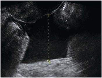
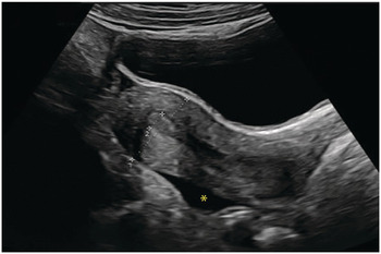
Ultrasound: multiple solid masses in the pouch of Douglas (23 mm ×... |  Download Scientific Diagram
Ultrasound: multiple solid masses in the pouch of Douglas (23 mm ×... | Download Scientific Diagram

Pouch of Douglas Liposarcoma—A Rarity

Ultrasound: multiple solid masses in the pouch of Douglas (23 mm ×... |  Download Scientific Diagram
Ultrasound: multiple solid masses in the pouch of Douglas (23 mm ×... | Download Scientific Diagram

Ultrasound, macroscopic and histological features of malignant ovarian  tumors. Metastatic tumors to the ovary: ovarian metastase
Ultrasound, macroscopic and histological features of malignant ovarian tumors. Metastatic tumors to the ovary: ovarian metastase

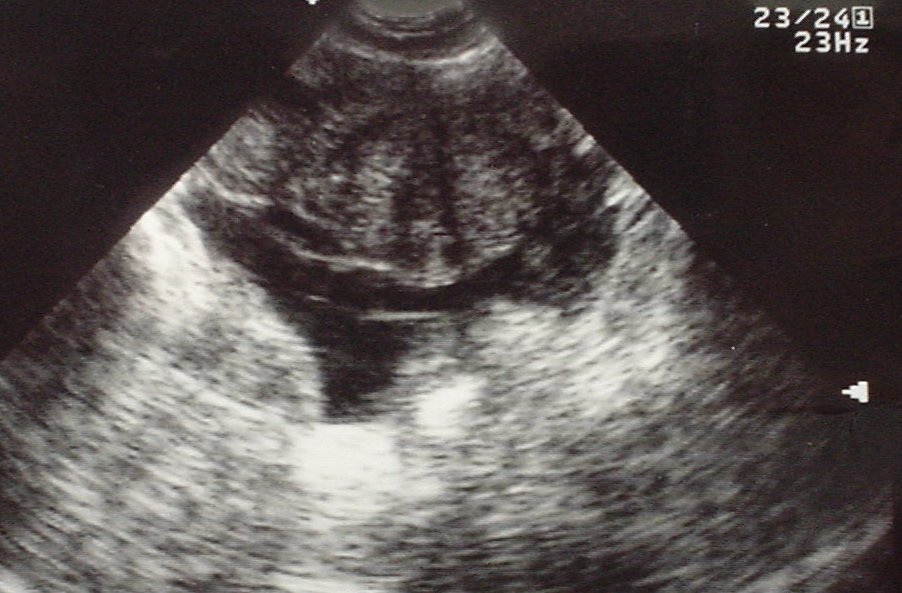
Gynaecology and Early Pregnancy Ultrasound (Chapter 8) - Ultrasound for the  Generalist
Gynaecology and Early Pregnancy Ultrasound (Chapter 8) - Ultrasound for the Generalist

Differential Diagnosis for Female Pelvic Masses | IntechOpen

Ultrasound: multiple solid masses in the pouch of Douglas (23 mm ×... |  Download Scientific Diagram
Ultrasound: multiple solid masses in the pouch of Douglas (23 mm ×... | Download Scientific Diagram

MRI of Tumors and Tumor Mimics in the Female Pelvis: Anatomic Pelvic  Space–based Approach | RadioGraphics
MRI of Tumors and Tumor Mimics in the Female Pelvis: Anatomic Pelvic Space–based Approach | RadioGraphics
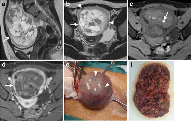
MR imaging of ovarian masses: classification and differential diagnosis |  Insights into Imaging | Full Text
MR imaging of ovarian masses: classification and differential diagnosis | Insights into Imaging | Full Text
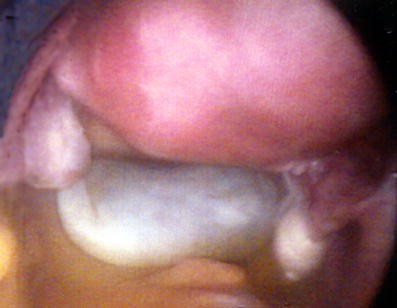
Ovarian torsion in a premenarchal patient | Eurorad

Ultrasound: multiple solid masses in the pouch of Douglas (23 mm ×... |  Download Scientific Diagram
Ultrasound: multiple solid masses in the pouch of Douglas (23 mm ×... | Download Scientific Diagram

Tomography | Free Full-Text | Mimics and Pitfalls of Primary Ovarian  Malignancy Imaging
Tomography | Free Full-Text | Mimics and Pitfalls of Primary Ovarian Malignancy Imaging

EPOS™
EPOS™

Ovarian torsion in a premenarchal patient | Eurorad

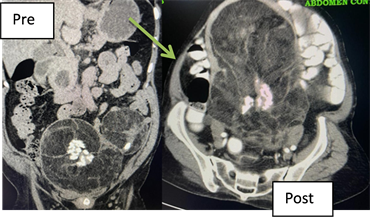
CT Abdomen and Pelvis – intravenous contrast showing large... | Download  Scientific Diagram
CT Abdomen and Pelvis – intravenous contrast showing large... | Download Scientific Diagram

Tomography | Free Full-Text | Mimics and Pitfalls of Primary Ovarian  Malignancy Imaging
Tomography | Free Full-Text | Mimics and Pitfalls of Primary Ovarian Malignancy Imaging

Pelvic Mass: Role of Imaging in the Diagnosis and Management | SpringerLink

Gynaecology and Early Pregnancy Ultrasound (Chapter 8) - Ultrasound for the  Generalist
Gynaecology and Early Pregnancy Ultrasound (Chapter 8) - Ultrasound for the Generalist

Nodular carcinomatosis on the peritoneum in the pouch of Douglas. |  Download Scientific Diagram
Nodular carcinomatosis on the peritoneum in the pouch of Douglas. | Download Scientific Diagram

EPOS™
EPOS™

Ultrasound: multiple solid masses in the pouch of Douglas (23 mm ×... |  Download Scientific Diagram
Ultrasound: multiple solid masses in the pouch of Douglas (23 mm ×... | Download Scientific Diagram

Benign cystic mesothelioma presenting as a solitary non–attached cyst in  the pouch of Douglas | Gynecological Surgery | Full Text
Benign cystic mesothelioma presenting as a solitary non–attached cyst in the pouch of Douglas | Gynecological Surgery | Full Text

Uncommon pelvic mass: GIST mimicking adnexal tumour | Eurorad

Gynecologic Ultrasound | SpringerLink

Primary and secondary tumors of the peritoneum: key imaging features and  differential diagnosis with surgical and pathological correlation |  Insights into Imaging | Full Text
Primary and secondary tumors of the peritoneum: key imaging features and differential diagnosis with surgical and pathological correlation | Insights into Imaging | Full Text

Pelvic Pain Tags - Differential Diagnosis of